Where are EEG electrodes placed?
The T3, C3, Cz, C4, and T4 electrodes are placed at marks made at intervals of 10%, 20%, 20%, 20%, 20% and 10%, respectively, measured across the top of the head. Skull circumference is measured just above the ears (T3 and T4), just above the bridge of the nose (at Fpz), and just above the occipital point (at Oz).
What are invasive electrodes?
When seizures can’t be easily localized, “invasive electrodes” are placed under the skull and on the surface of the brain or into certain brain areas. There are two main types of invasive electrodes: strips or grids and depth electrodes. These electrodes can pick up brain activity much better than scalp electrodes.
Which electrode is used to detect brain activity?
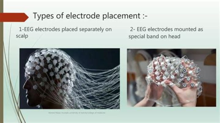
An EEG is a test that detects abnormalities in your brain waves, or in the electrical activity of your brain. During the procedure, electrodes consisting of small metal discs with thin wires are pasted onto your scalp. The electrodes detect tiny electrical charges that result from the activity of your brain cells.
How many electrodes does an EEG have?
An array of 25 electrodes is recommended for standard EEGs with inferior temporal electrodes. Due to thinner skulls (spatial aliasing), pediatric EEG requires as many scalp electrodes as in adults. Arrays with higher electrode numbers (64–256 electrodes) allow source imaging at sublobar level.
What are the four basic EEG patterns?
Four simple periodic rhythms recorded in the EEG are alpha, beta, delta, and theta. These rhythms are identified by frequency (Hz or cycles/sec) and amplitude (Table 3.1).
Are EEGS invasive?
An electroencephalogram (EEG) is the recording of the brain electrical activity. A set of electrodes are placed on the scalp of the subject. This technique is non-invasive since no surgery is required. EEG is a fast and cheap technique.
What is the size of the depth electrodes?
The standard subdural contact electrodes are 2–3 mm in diameter and separated by 5–10 mm. Microgrids have electrode contacts that are 1.5 mm in diameter and separated by 4 mm. Both are placed through a craniotomy with a size depending on the extent of cerebral surface that is to be covered.
How many electrodes are used in a 12 lead ECG?
Although it is called a 12-lead ECG, it uses only 10 electrodes. Certain electrodes are part of two pairs and thus provide two leads. Electrodes typically are self-adhesive pads with a conducting gel in the centre.
How does a normal EEG look?
Normal EEG waveforms, like many kinds of waveforms, are defined and described by their frequency, amplitude, and location. Frequency (Hertz, Hz) is a key characteristic used to define normal or abnormal EEG rhythms. Most waves of 8 Hz and higher frequencies are normal findings in the EEG of an awake adult.
Which brain waves are most Desynchronous?
When awake, most people exhibit brain wave, (EEG) patterns that can be classified into two types of waves, beta and alpha. Beta waves are those associated with day to day wakefulness. These waves are the highest in frequency and lowest in amplitude, and also more desynchronous than other waves.
Is ERP invasive or non-invasive?
Invasiveness. Unlike microelectrodes, which require an electrode to be inserted into the brain, and PET scans that expose humans to radiation, ERPs use EEG, a non-invasive procedure.
Why does EEG have poor spatial resolution?
Here, we argue that the actual temporal resolution of conventional (scalp potentials) EEG is overestimated, and that volume conduction, the main cause of the poor spatial resolution of EEG, also distorts the recovered time course of the underlying sources at scalp level, and hence degrades the actual temporal …
How are depth electrodes placed?
Subdural EEG electrodes are those electrodes which sit over the surface of the brain. Depth EEG electrodes are those electrodes which are placed within the substance of the brain.
What are depth electrodes made of?
The depth electrodes were made of either platinum or a nickel-chromium alloy (nichrome). We reviewed 98 cases in which patients with implanted depth electrodes, subdural electrodes, or both underwent MR scanning.
Why is 12 lead ECG called 12?
The 12-lead ECG displays, as the name implies, 12 leads which are derived by means of 10 electrodes. Three of these leads are easy to understand, since they are simply the result of comparing electrical potentials recorded by two electrodes; one electrode is exploring, while the other is a reference electrode.
What are the 12 ECG leads?
A 12-lead ECG consists of three bipolar limb leads (I, II, and III), the unipolar limb leads (AVR, AVL, and AVF), and six unipolar chest leads, also called precordial or V leads, ( , , , , , and ).